From Wikipedia, the free encyclopedia
Buruli ulcer also known as Biarnsdale ulcer is an infectious disease caused by bacteria. These bacteria are related to the ones that cause leprosy and those that cause tuberculosis. The disease can be found in tropical regions, but is not limited to the tropics. It is common in Africa, Southeast Asia and Australia. Currently, it is unknown in what way the disease spreads. It often occurs in rural areas close to swamps, rivers, or lakes. For this reason, vector organisms such as mosquitoes appear likely.
Locations of Buruli ulcer outbreaks in Australia
Buruli ulcer has been reported in 33 countries around the world. Affected areas include rural West Africa, Central Africa, New Guinea, Latin America and tropical regions of Asia.
In Australia, Buruli ulcer most commonly occurs in localised coastal areas of Victoria. It was first diagnosed in the Bairnsdale area of East Gippsland in the 1930s. Since then, cases have been reported from Western Port and Phillip Island, the Frankston–Seaford area, the Bellarine Peninsula, the Mornington Peninsula and other bayside areas in south eastern Melbourne. Buruli ulcer also occurs less commonly in the Daintree region of Far North Queensland.
Symptoms of Buruli ulcer
The progression of symptoms can include:
A spot that looks like a mosquito or spider bite forms on the skin (most commonly on the limbs).
The spot grows bigger over days or weeks.
The spot may form a crusty, non-healing scab.
The scab then disintegrates into an ulcer.
The ulcer continues to enlarge.
Unlike other ulcers, this ulcer is usually painless and there is generally no fever or other signs of infection.
The infection may sometimes present with no ulceration but with localised pain, swelling and fever, raised lumps, or thickened or raised flat areas of skin.
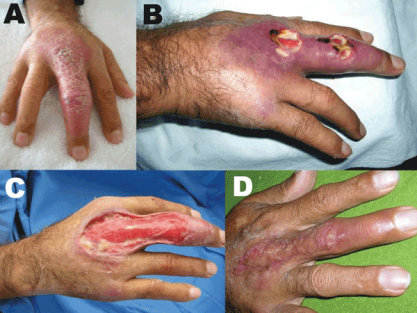
Buruli ulcer on the hand of a person from Peru.
A) nonulcerative edematous lesion on the right middle finger as first seen
B) ulcerated lesions on the right middle finger about 4 weeks later
C) extensive debridement, 5.5 weeks after first seen
D) cured lesion 5 months after first seen, 1 month after autologous skin graft
Diagnosis
The diagnosis of Buruli ulcer is usually based on the characteristic appearance of the ulcer in an endemic area. If there is any doubt about the diagnosis, then PCR using the IS2404 target is helpful, but this is not specific for M. ulcerans. The Ziehl-Neelsen stain is only 40–80% sensitive, and culture is 20–60% sensitive. Simultaneous use of multiple methods may be necessary to make the diagnosis.
Prevention
There is no specific vaccine for Myocobacterium ulcerans. The Bacillus Calmette-Guérin vaccine may offer temporary protection.
Treatment
If treated early, antibiotics for eight weeks are effective in 80% of people. Treatment may also include cutting out the ulcer. This may be a minor operation and very successful if undertaken early. Advanced disease may require prolonged treatment with extensive skin grafting. Surgical practice can be dangerous in the developing countries where the disease is common.
There are different forms of thearpy: the most common one consists of a combination two or three different drugs, and removing the affected tissue. Simple tissue removal looks promising, when no ulcers have formed yet.[1] The bacterium does not support temperatures above 40 °C. For this reason, some forms of therapy have included heating the affected skin parts above 40°C. This will kill the bacteria. First attempts suggest that after killing the bacteria, the ulcer will heal in several cases. Even 18 months after the treatment, the disease no longer shows. This means that the method can be tested on more people.
History
James Augustus Grant, in his book A Walk across Africa (1864), describes how his leg became grossly swollen and stiff with later a copious discharge. This was almost certainly the severe edematous form of the disease, and is the first known description of the infection[citation needed]. Buruli ulcer disease was identified in 1897 by Sir Albert Cook, a British physician, at Mengo Hospital in Kampala, Uganda. A detailed description of the disease was written in 1948 by Professor Peter MacCallum and his colleagues, who were treating patients from the Bairnsdale district, near Melbourne, Australia. They were the first to identify Mycobacterium ulcerans as the pathogen causing it. The disease was so named after Buruli County in Uganda (now called Nakasongola District), because of the many cases that occurred there in the 1960s. In Australia it is also known as Bairnsdale or Daintree ulcer. The incidence of the disease has recently been rising in tropical Africa and in certain parts of Australia.
In March 2008, researchers announced the first isolation of M. ulcerans from the environment. This suggested that the disease might be transmitted via contact with the environment rather than person to person. The entire genome of M. ulcerans has been sequenced.
Certain types of clay have historically been used in an attempt to treat the condition.